Background
Biocytogen provides preclinical assessment services for gene therapy products, which involve the transfer of genetic material to treat or prevent disease. This process changes the way cells produce proteins or groups of proteins, allowing us to reduce levels of disease-causing versions of proteins, increase production of disease-resistant proteins, or introduce new/modified genes into the body to help treat a disease. Our services are designed to ensure that gene therapy products meet safety and efficacy standards before they enter clinical trials.
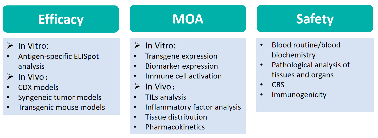
Our pharmacology platform is designed to evaluate the efficacy, safety and mechanism of action of gene therapy products in support of Investigational New Drug (IND) applications. Our flow cytometry platform and cell base platform support a series of in-vitro and ex-vivo studies in a quick, reliable, and reproducible way, including the evaluation of immune cell activation, tissue distribution, immunogenicity etc. Additionally, our full-fledged team with abundant animal model resource efficiently support the efficacy evaluation of gene therapy products in vivo. Furthermore, our pathology/toxicology platform provides comprehensive drug safety evaluation during preclinical study.
Results
Animal models

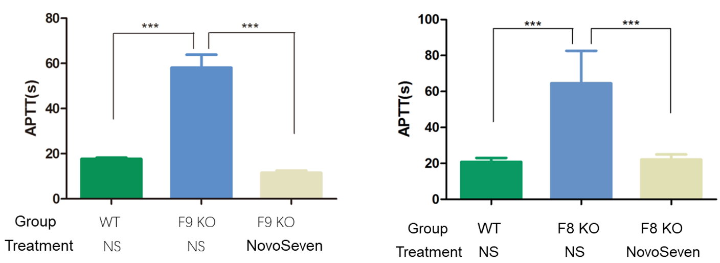
Fig1. Activated partial thromboplastin time assay. The results showed that APTT values in F9/F8 KO mice were much higher than those in WT mice, and APTT returned to normal values after injection of NovoSeven.
Syngeneic tumor models & CDX models
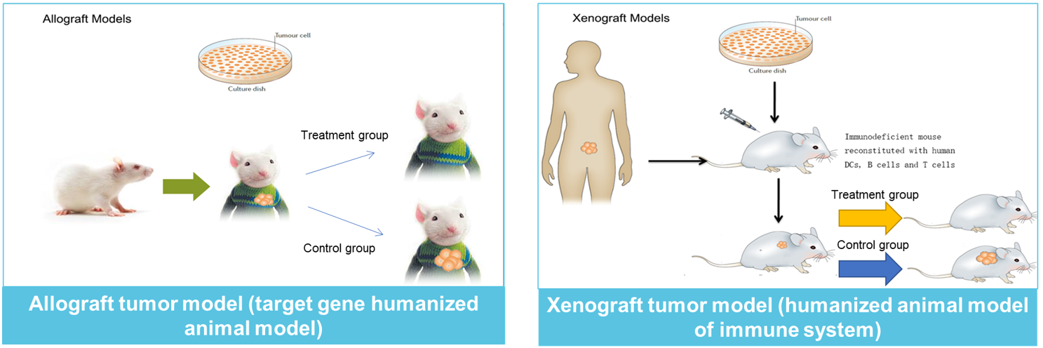
ELISpot assay


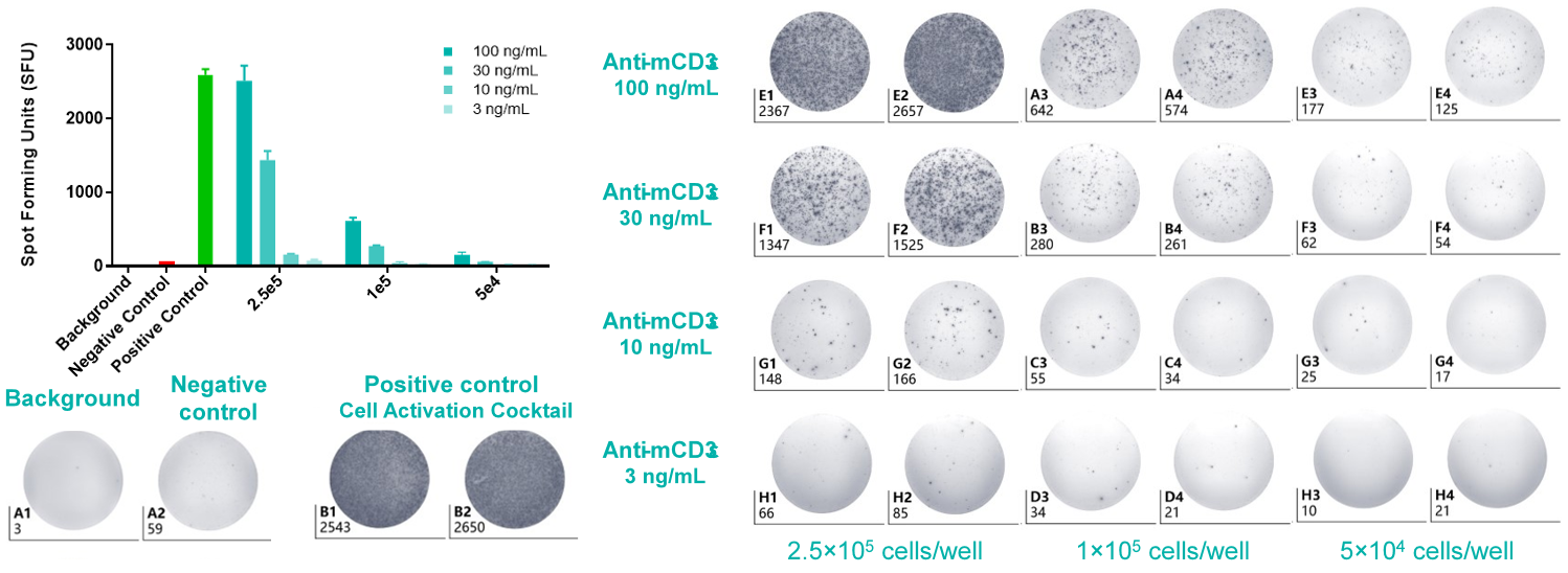
Fig2. IFN-γ ELISpot assay of splenocytes in response to stimulation with an mCD3e antibody. The statistical histogram and plate view of IFN-γ ELISpot assay. Splenocytes from C57BL/6 mice were seeded at density at 5.00×10⁴-2.50×10⁵/well. The number of spots indicates how much IFN-γ was produced by the splenocytes in response to stimulation with mCD3e antibody, providing information about immune cell activation levels.

Fig3. Flow Cytometry Analysis of Intracellular proteins. Tumor infiltrating lymphocytes (TILs) were isolated from MC38 tumor inoculated in C57BL/6 mice. TNF-α/IFN-γ in tumor-infiltrating T cells and Granzyme B in tumor-infiltrating T/NK cells were measured by FACS.

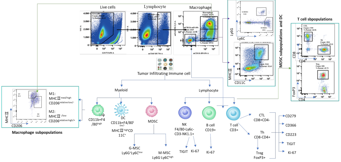
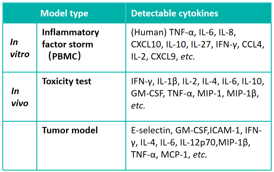
Fig4. Immune-profiling by Flow Cytometry in a single panel. TNF-α/IFN-γ in tumor-infiltrating T cells and Granzyme B in tumor-infiltrating T/NK cells.

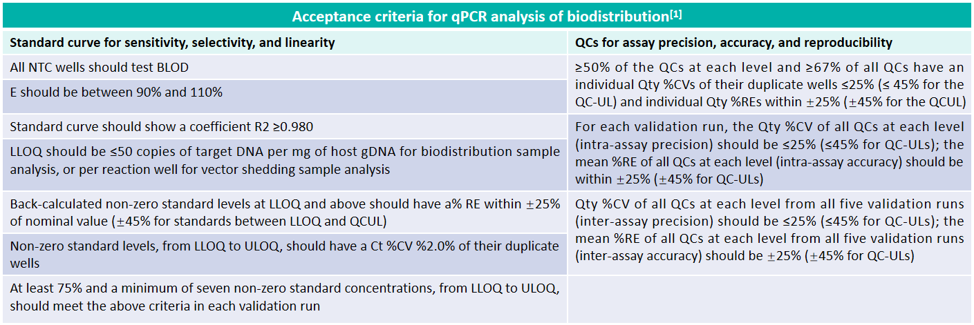
*RT-qPCR assay is undergoing method development and validation
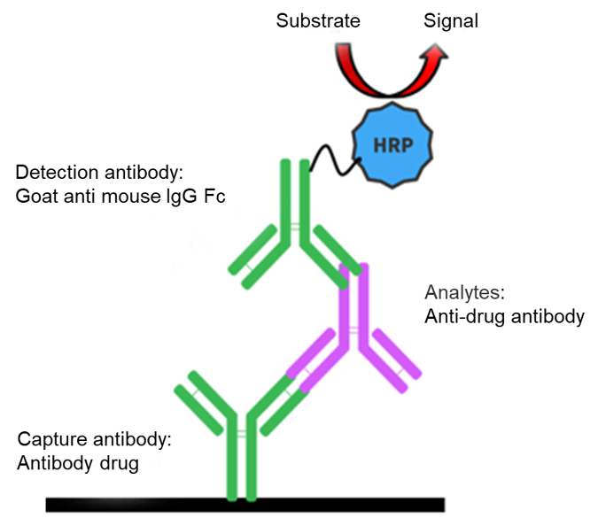
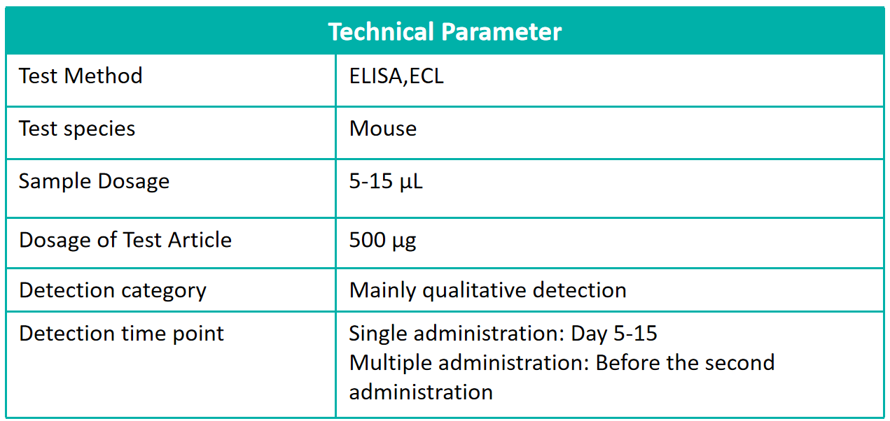
ADA assay
Pathology/Toxicology
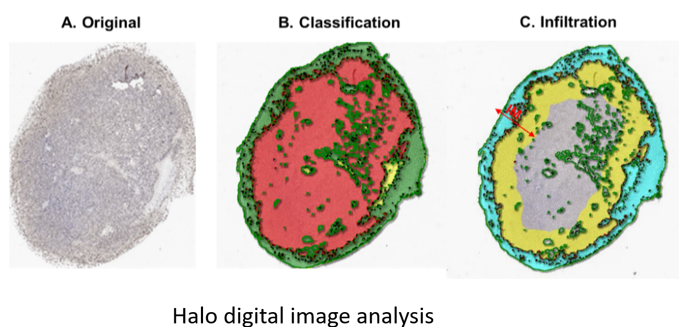
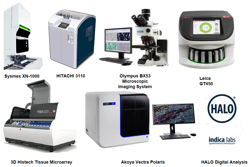
Anatomic pathology: H&E, IHC, IF, ICC & special stains
Molecular pathology: RNAscope & Tunel staining
Diagnostic pathology
Toxicological evaluation
Qualitative & Semi-quantitative analyses
Tissue microarray drug screening
Clinical haematology: CBC & biochemistry
Reference
[1] Ma H, Bell KN, Loker RN. Mol Ther Methods Clin Dev. 2020 Nov 17;20:152-168.


 +86-10-56967680
 info@bbctg.com.cn