Delayed-type hypersensitivity (DTH) is an inflammatory response characterized by mononuclear cell infiltration and tissue cell damage after the action of effector T cells with corresponding antigens[1]. It usually occurs during bacterial and viral infections, oncological diseases, transplant rejection, and contact dermatitis.
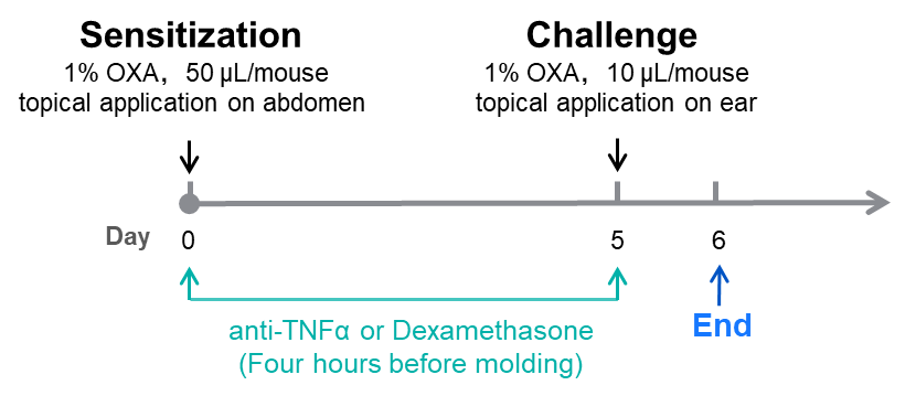
Mouse contact hypersensitivity response is a widely used model to study T cell-mediated immune responses to contact allergens. Oxazolone (OXA) is a hapten, which can combine with skin proteins to form complete antigens when applied to the skin, stimulating the proliferation of T lymphocytes into sensitized lymphocytes[2]. When applied again with OXA after 4-7 days, it causes a delayed allergic reaction locally.
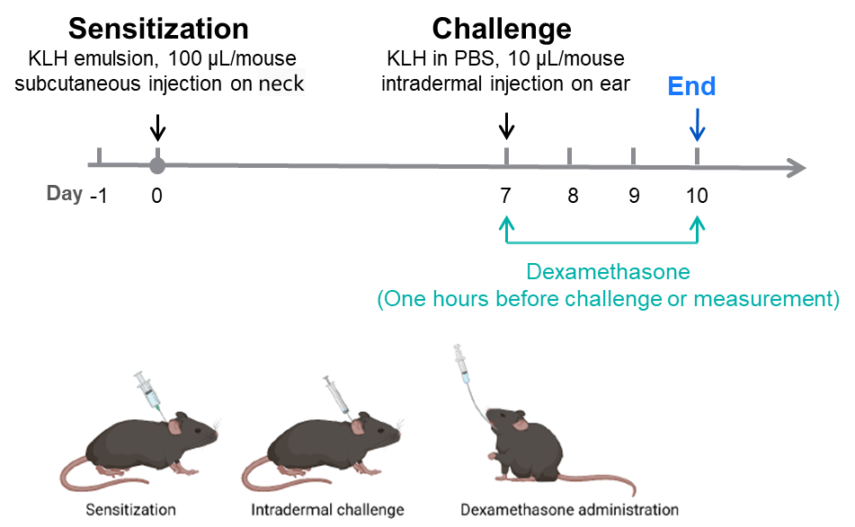
Keyhole limpet hemocyanin (KLH) is a highly immunogenic protein macromolecule that enables dendritic cells to recognize Th1 cells[3]. It is an ideal antigen for inducing late DTH. KLH is introduced systemically in mice. And then locally reintroduced several weeks later, DTH response was assessed by relative swelling at the injection site.
OXA and KLH-induced DTH mouse (Balb/c) models were established by Biocytogen, which can be used for immunosuppressant efficacy evaluation.
OXA-Induced DTH Mouse Model based on BALB/c Mice

KLH-Induced DTH Mouse Model based on C57BL/6 Mice

Efficacy Evaluation of Dexamethasone and Anti-TNFα in a OXA-induced DTH Mouse Model


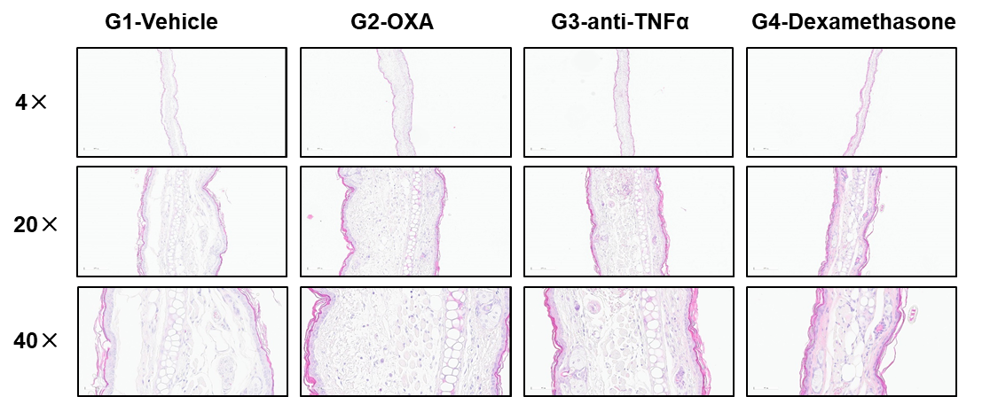
Figure 2. Histologic Assessment of DTH in BALB/c Mice
Efficacy Evaluation of Dexamethasone on KLH-induced DTH model

Figure 3. Dexamethasone alleviates KLH-induced an DTH phenotype in C57BL/6 mice. Body weight (A), (B) Ear Thickness. Values are expressed as mean ± SEM.

References
[1] Honda T, Egawa G, Grabbe S, Kabashima K. Update of immune events in the murine contact hypersensitivity model: toward the understanding of allergic contact dermatitis. J Invest Dermatol. 2013. 133(2):303-15.
[2] Mchale J F , Harari O A , Marshall D , et al. Vascular endothelial cell expression of ICAM-1 and VCAM-1 at the onset of eliciting contact hypersensitivity in mice: evidence for a dominant role of TNF-alpha.[J]. American Association of Immunologists, 1999(3).
[3] Richerson HB, Dvorak HF, Leskowitz S. Cutaneous basophil hypersensitivity. I. A new look at the Jones-Mote reaction, general characteristics. J Exp Med. 1970. 132(3):546-57.


 +86-10-56967680
 info@bbctg.com.cn