Osteoporosis has been defined as a systemic skeletal disease that is characterized by low bone mass and micro-architectural deterioration of bone tissue, with a consequent increase in bone fragility and susceptibility to fracture [1]. The basic pathogenesis of osteoporosis is deregulation of bone formation and resorption. Ovariectomized mice are an accepted in vivo model of human Postmenopausal osteoporosis (PMOP). Bone mineral density (BMD) measurement, micro-CT analysis and histomorphometric analysis were used to evaluate the OVX mice [2, 3]. Bones were harvested and tested after the operation.
A stable OVX-induced osteoporosis disease model protocol was established in C57BL/6 mice by Biocytogen, which can be used for preclinical studies and pharmacodynamic evaluation of osteoporosis.
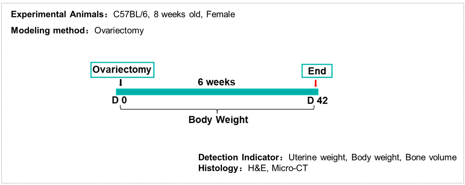
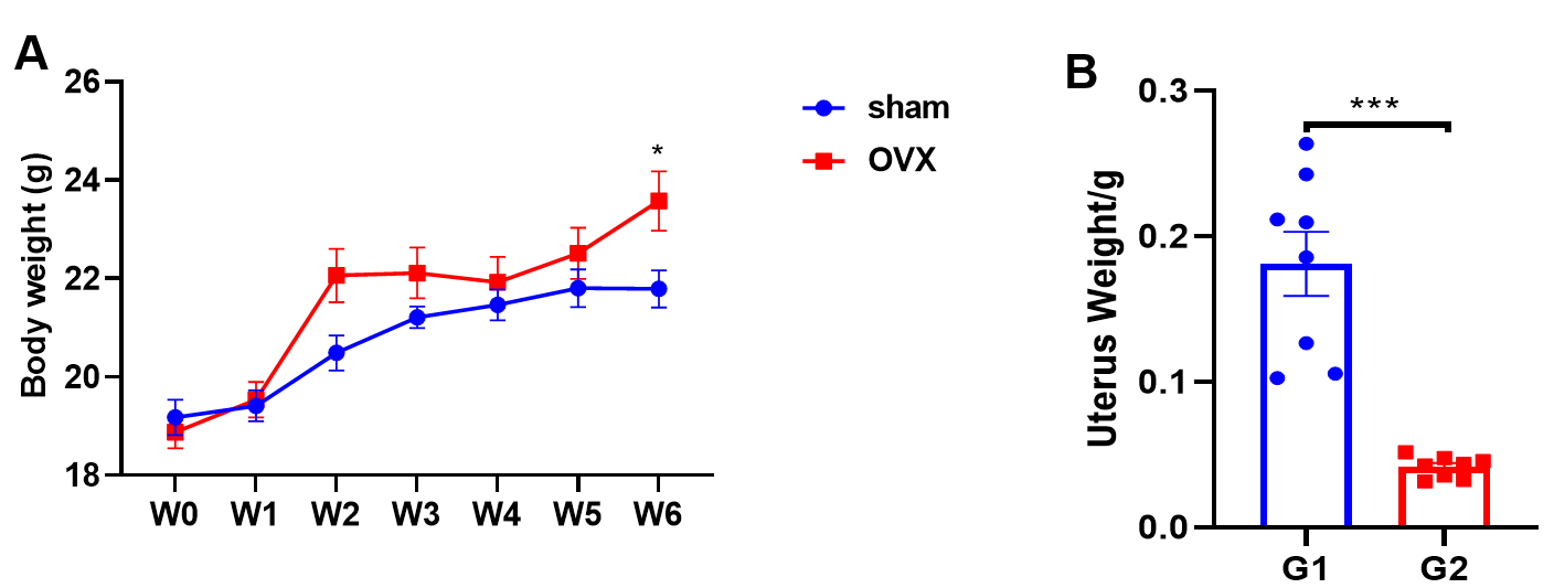
OVX induced weight loss of body and uterus in mice
OVX induced bone loss in mice (CT)
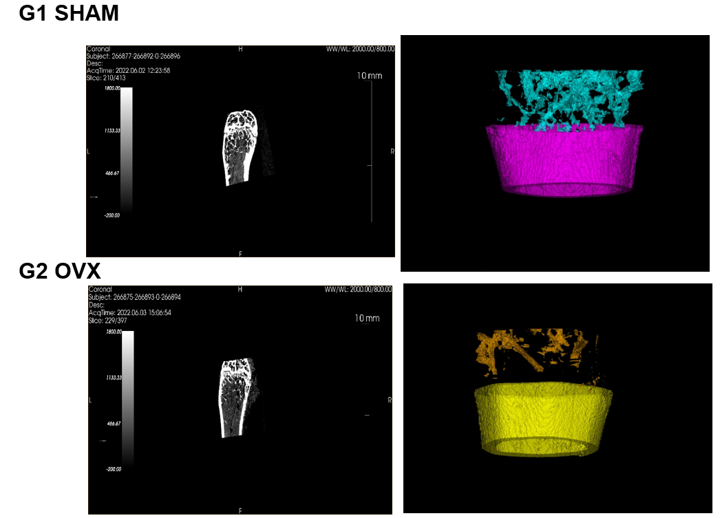
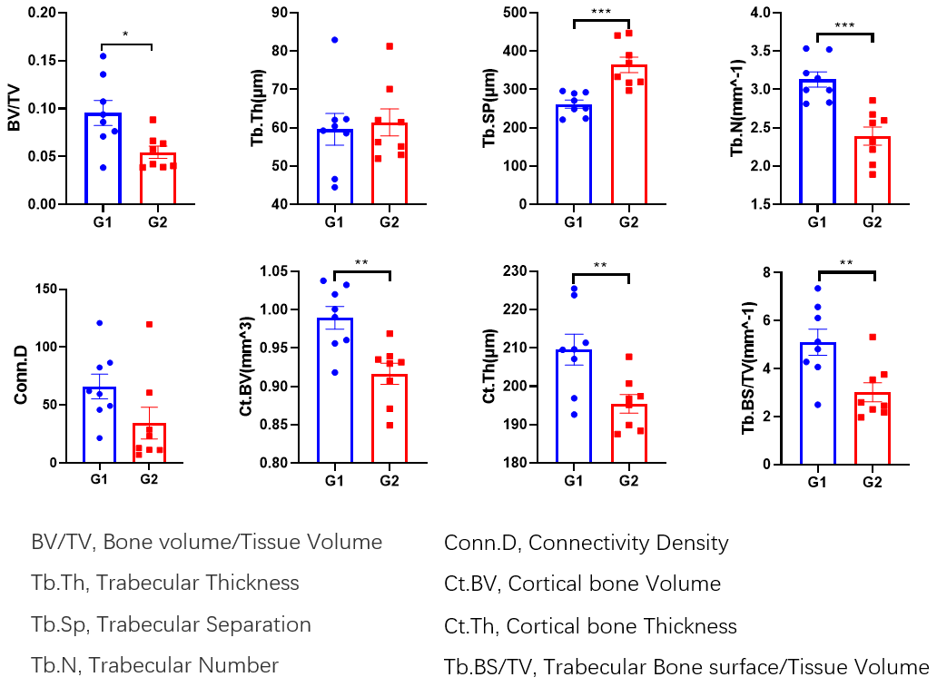
Ovariectomy induced osteoporosis. C57BL/6 mice received ovariectomy on day 0. Six weeks after operation, bones and uterus were harvested and tested. Body weight were recorded everyday. Ovariectomy produced a bone loss in mice. Values are expressed as mean ± SEM. n=8, T-test, * p<0.05, **p<0.01.
OVX induced bone loss in mice (H&E)
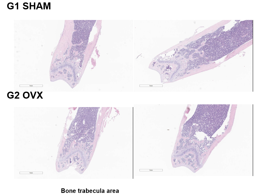
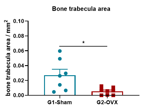
Pathological analysis of ovariectomy-induced osteoporosis. Six weeks after ovariectomy, the bone trabecula area was decreased in mice that received OVX surgery. Values are expressed Values are expressed as mean ± SEM. n=8, T-test, * p<0.05, **p<0.01.

References
1. Eastell R, O‘Neill TW , Hofbauer LC et al. Postmenopausal osteoporosis. Nat Rev Dis Primers. 2016, 2:16069.
2. Wang HC, Zhou KF, Xiao FZ et al. Identification of circRNA-associated ceRNA network in BMSCs of OVX models for postmenopausal osteoporosis. Sci Rep. 2020, 10(1):10896.
3. Chen K, Qiu PC, Yuan Y et al. Pseurotin A Inhibits Osteoclastogenesis and Prevents Ovariectomized-Induced Bone Loss by Suppressing Reactive Oxygen Species. Theranostics. 2019, 9(6):1634-1650.


 +86-10-56967680
 info@bbctg.com.cn